Introduction to Food Microbiology and Safety
Gram Staining: Principles, Methods, and Procedures
Gram staining is a cornerstone technique in bacteriology, widely used to classify bacteria into Gram-positive and Gram-negative groups based on cell wall structure. In this article, we provide a concise overview of its principles, step-by-step procedures, and interpretation of results. While this guide is aimed at beginners in food microbiology, it serves as a practical resource for understanding the fundamental aspects of Gram staining without delving into exhaustive experimental details.
Principles of Gram Staining
Gram staining is a fundamental bacteriological technique, particularly relevant in food microbiology. It differentiates bacteria into Gram-positive and Gram-negative groups based on the thickness and composition of their cell walls. The process involves a series of staining and decolorization steps that highlight these structural differences.
Procedure
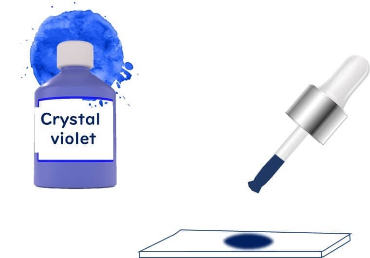
Crystal Violet Staining
The procedure begins by staining bacterial cells with crystal violet, a purple dye that adheres to cell walls. At this stage, both Gram-positive and Gram-negative bacteria appear purple.
However, Gram-negative bacteria, with their thinner cell walls, absorb the dye less intensely.
Iodine Fixation
Next, iodine solution (Gram's iodine) is applied, forming a complex with crystal violet in Gram-positive bacteria.
This step ensures that the dye is fixed firmly to the thick peptidoglycan layer of Gram-positive bacteria.
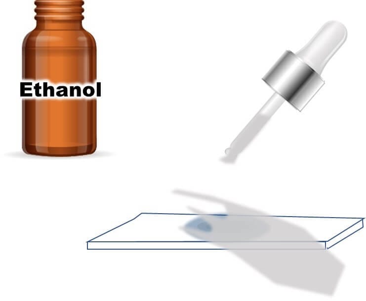
Decolorization
Alcohol is used to decolorize the cells.
Gram-positive bacteria retain the purple dye due to their thick peptidoglycan layer, while Gram-negative bacteria, with their thinner walls, lose the dye.
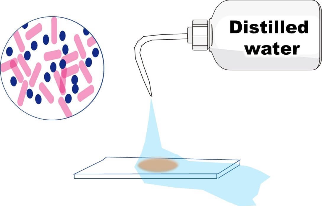
Counterstaining with Safranin
To differentiate further, safranin, a pink counterstain, is applied.
Gram-negative bacteria, having lost the crystal violet, take up the pink safranin, while Gram-positive bacteria retain their purple color.
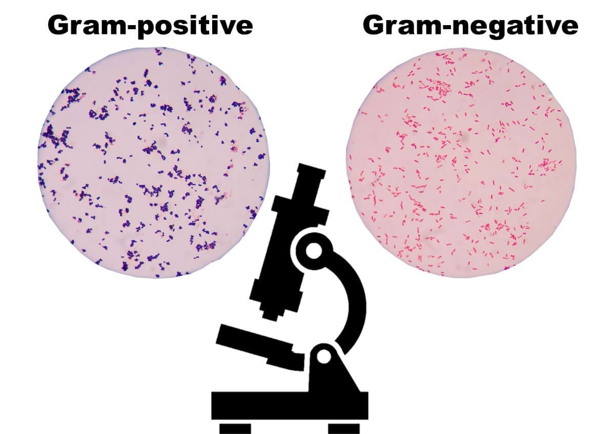
Results
Gram-positive bacteria: Appear purple due to retention of crystal violet.
Gram-negative bacteria: Appear pink as they absorb the counterstain safranin.
This color distinction forms the basis for identifying and categorizing bacteria in food microbiology and other bacteriological applications.

Discover expert-led lessons in food microbiology designed for professionals and beginners alike.

Author of this Blog: Bon Kimura
Bon Kimura, Professor Emeritus at Tokyo University of Marine Science and Technology (TUMSAT), specializes in food microbiology. He obtained his PhD from Kyoto University and became a professor at TUMSAT in 2006, serving as Dean of the Faculty of Marine Science from 2012-2015. Kimura has published over 200 international papers on food safety, pathogens, and spoilage bacteria. He has received multiple awards, including the Japanese Society for Food Microbiology Award (2019). Kimura also served as an editor for the International Journal of Food Microbiology from 2012 to 2024, where he was the principal reviewer for 1,927 papers.
Copyright © Introduction to Food Microbiology and Safety All Rights Reserved.